b value mri
Computed DWI images up to a simulated b. The degree of diffusion weight in g correlates with the strength of the diffusion gradients characterized b y the b - value which is a function of the gradient related parameters.

Differential Tractography Dsi Studio Documentation
A baseline b-value of 50 smm² is often used in liver diffusion-weighted imaging instead of b 0.
. Units are micro-Tesla µT. The b value is used in MRI in the context of Diffusion Weighted Imaging DWI. DWI is done to determine the rate of molecular diffusion in different areas of the body.
MATERIALS AND METHODS Patients. Certa in illnesses show restrictions of diffusion for example demyel in ization and cytotoxic edema. We found significant differences in the qualitative points values among the DW images with different b values F 30218 p 0001.
In general in healthy tissue molecules of water and other chemicals are not stationary but moving about. Fortytwo consecutive patients underwent multi bvalue 16 evenly spaced bvalues between 0 and 2000 smm 2 DWI along with multiparametric MRI MPMRI of the prostate at 3 Tesla followed by transrectal ultrasoundMRI fusion guided targeted biopsy of suspicious lesions detected at MPMRI. The proper b-value has approximately 80 of the reciprocal ADC value of nor- mal background tissue.
Also may be called the turbo factor. High b-value diffusion-weighted MRI of normal brain Brain DW images obtained at b 3000 appear significantly different from those obtained at b 1000 reflecting expected loss of signal from all areas of brain in proportion to their ADC values. Smaller b values correlated with better DW image quality.
There is no consensus on the optimal b-value. B1rms The root-mean-square value of the MRI effective component of the B1 field. Modern scanners usually allow a range of 0-4000smm 2.
The reason is readily apparent from the images below. Two recent studies explored DWI at an ultra high b-value of 2000smm 2. Lesions were analyzed for benignitymalignity using apparent diffusion coefficient ADC values with 10 b-value combinations and by measuring the lesionnormal parenchyma ADC ratio.
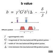
B γ² G² δ² Δδ3. Diffusion magnetic resonance imaging dMRI is a unique technique to probe the microstructure of the normal and diseased tissue by quantifying the displacements of water molecules 1. With b 0 bright signals are noted in multiple veins due to the high T2 of blood coupled with sluggish flow.
The use of b values more than 1000 smm 2 would offer better contrast but was more liable to suffer susceptibility artifact. Strength duration and the period b etween diffusion gradients. A b value of 8001000 smm 2 would provide an excellent spatial resolution and an adequate signalnoise ratio for lesion evaluation.
This can be compensated by increasing averages which result in longer scan times. The brain was scanned on a slice of the basal nucleus in a direction parallel to the anterior commissureposterior commissure line. DBS Deep brain stimulation.
Depending on the organ being imaged b-values typically range from 50-1000smm 2. The images acquired with low b values are less diffusion-weighted because they use less gradient. The mean SNR of the images ranged from 2175 364 at a b value of 0 smm2 to 531 317 at a b value of 800 smm2.
B1 The RF magnetic field produced by the MRI scanner. The aim of our study was therefore to determine with the aid of histopathological findings for radical prostatectomy as a reference which bvalue b 1000 smm 2 or b 2000 smm 2 used for 3T MRI is more suitable for visual assessment for the detection of prostate cancer on native DWI. In DWI we recommend the use of b-values of 0 and 800 smm 2 as two b-values or b0 50 600 800 and 1000 smm 2 as multiple b-values for distinguishing between benign and malignant liver lesions.
On clinical MRI scanners diffusion sensitivity is easily altered by changing the parameter known as the b value which is mainly proportional to the gradient amplitude and duration 17. Most prior diffusion-weighted imaging studies of human brain infarction have been performed with b values of 1000 or less 2126 although one reported a b value of 1463 13. Keep in mind that higher b-values may pronounce lesions even more at the price of poor SNR due to longer TEs and increased susceptibility.
Mean ADC value is 13 higher in total by additional use of b0 and b50 smm 2 in multiple b-value combinations. The purpose of this study was to determine the role of high-b-value b 2500 or 3000 diffusion-weighted imaging for lesion detection in acute and chronic brain infarction. Units are micro-Tesla µT.
The motion probing gradients were applied in three orthogonal directions at b values of 0 300 500 700 1000 1300 1500 1800 2000 2300 2500 2800 3000 3500 4000 and 5000 smm 2. B value measures the degree of diffusion weighting applied thereby indicating the amplitude G time of applied gradients δ and duration between the paired gradients Δ and is calculated as. ETL Echo train length.
Routine abdominal MRI and DWI were performed using seven b-values 0 50 200 400 600 800 1000 smm 2. At b 3000 smm 2 points from 1 to 3 were assigned to tumor signals the same as those observed at b 1000 smm 2 but 4 points were assigned to hyperintense signals between those of the normal subcortical white matter and the corticospinal tract and 5 to markedly hyperintense signals that were equal to or higher than that of the normal. The term b -value characterizes the diffusion gradient pulses amplitude shape and timing and expresses the amount of diffusion weighting 1.

Principles Of Diffusion Tensor Imaging And Its Applications To Basic Neuroscience Research Neuron

Apparent Diffusion Coefficient Radiology Reference Article Radiopaedia Org
Tensor Valued Diffusion Encoding For Diffusional Variance Decomposition Divide Technical Feasibility In Clinical Mri Systems Plos One
2
2

Principles Of Diffusion Tensor Imaging And Its Applications To Basic Neuroscience Research Neuron
2
Diffusion Weighted Imaging Radiology Reference Article Radiopaedia Org

Diffusion Weighted Imaging Radiology Reference Article Radiopaedia Org

Signal Intensity Of Dwi And Adc In Diffusion Restriction Increased Download Scientific Diagram
2
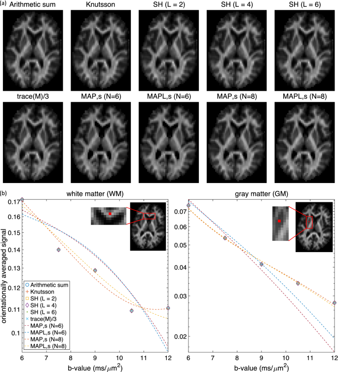
Computing The Orientational Average Of Diffusion Weighted Mri Signals A Comparison Of Different Techniques Scientific Reports

A Diffusion Mri Based Spatiotemporal Continuum Of The Embryonic Mouse Brain For Probing Gene Neuroanatomy Connections Pnas

Hyperpolarised 13c Mri Identifies The Emergence Of A Glycolytic Cell Population Within Intermediate Risk Human Prostate Cancer Nature Communications

Diffusion Tensor Imaging And Fiber Tractography Radiology Reference Article Radiopaedia Org
2

Apparent Diffusion Coefficient Radiology Reference Article Radiopaedia Org

Principles Of Diffusion Tensor Imaging And Its Applications To Basic Neuroscience Research Neuron
Tensor Valued Diffusion Encoding For Diffusional Variance Decomposition Divide Technical Feasibility In Clinical Mri Systems Plos One


Comments
Post a Comment